This was seen in this case during the intra-operative exploration. Īnterior dislocation may happen in a direct force to the dorsal aspect of forearm with the elbow in semi-flexed position, with avulsion of the tip of olecranon near the insertion of triceps. Anterior elbow dislocation is rare due to strong bony stabilising effect of ulno-humeral joint and soft tissue structure of the posterior column, namely the triceps and posterior capsule. The peak incidence of paediatric elbow dislocation is when physis begins to close between 10 to 15 years of age. Range of movement of patient’s left elbow at 8 weeks post-op.: a - flexion b - extension c - supination d - pronationĮlbow dislocation in paediatric age group is uncommon. The elbow was immobilised at 90° flexion with forearm in supination in a splint (Figure 2).įig. Brachial, radial and ulnar pulses were good after reduction and repair. Intra-operative stability assessment was done with elbow joint was found stable in supination, pronation, flexion and extension. Medial collateral ligament (MCL) was completely torn and was repaired using 5/0 absorbable suture. The fracture was fixed with two parallel K-wires and the radial head was reduced spontaneously. Gentle controlled traction was applied to the elbow for bony reduction. The olecranon was found fractured at its metaphysis with a sleeve of bone attached to its physeal-epiphyseal region. Ulnar nerve was mobilised proximally and distally until tension-free. It was stretched and appeared slightly pale but was in continuity. Intra-operatively ulnar nerve was found impinged by the distal ulnar fragment just distal to the medial condylar groove. Medial approach of the elbow was chosen for assess of ulna nerve exploration and fracture reduction. Open reduction and percutaneous K-wire insertion under general anaesthesia was performed approximately 6 hours post trauma.
#Left olecranon fracture manual#
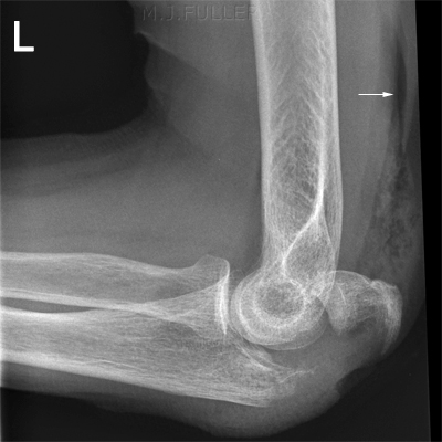
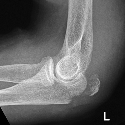
Close manual reduction was attempted approximately two hours post trauma at casualty but failed. The plain radiographs of her left elbow revealed anterior dislocation of the left elbow with olecranon fracture (Figure 1). However, the brachial and distal pulses were present. Ulnar claw was observed with reduced sensation over ulnar nerve distribution. Clinically, her left elbow was swollen and deformed. She was brought immediately to casualty by her parents. Clinical caseĪ healthy six-year-old girl sustained injury to her left elbow after she tripped and fell in the toilet. Authors report a case of anterior fracture dislocation of the elbow with ulnar nerve palsy in a six-year-old girl. Anterior elbow dislocation is rarely reported, with incidence of only <2%.

There were limited studies reported on paediatric elbow dislocation, which of most were case reports with rare type of dislocations and complications. Other subtypes which were less common include anterior, medial, lateral, convergent, and divergent dislocations. Of the reported cases to date, three-quarter were posterior dislocation of the elbow. Commonly, it is accompanied by concomitant fractures even though isolated dislocations have been reported. Traumatic elbow dislocation in children is rare, accounting for only 3–6% of paediatric elbow injuries. Surgical approach should be tailored individually according to the instability of the elbow joint and neurovascular status, as in this case was the posteromedial instability associated with ulnar nerve palsy. This rare injury should be treated with high index of suspicious. We chose medial approach of the elbow for ulnar nerve exploration and olecranon fixation.Ĭonclusion. There was no clear recommendation in the literature regarding surgical approach. Anterior elbow dislocation is a high energy trauma and one should be cautious of neurovascular injury. The elbow was stable in varus and valgus.ĭiscussion. At 8 weeks, range of movement of the elbow was full. The fracture heals and the K-wires were removed at 6 weeks.

Ulnar nerve was mobilised until tension-free. The transverse olecranon fracture was fixed with two K-wires and the radial head was reduced. Intra-operatively ulnar nerve was found impinged by the distal ulnar fragment but was in continuity. Open reduction and percutaneous K-wire insertion under general anaesthesia was performed. Closed manual reduction was attempted but failed. The X-rays showed anterior dislocation of the left elbow with olecranon fracture. No wound was found, distal pulses and circulation were good. Ulnar clawing was present with reduced sensation over ulnar nerve distribution. A 6-year-old girl presented to casualty with left elbow deformity and pain after she tripped and fell in the toilet. Anterior elbow dislocation is rarely reported, with incidence of only <2%.Ĭlinical case. Anterior elbow fracture dislocation is rare, especially in paediatric age group.


 0 kommentar(er)
